
Introduction
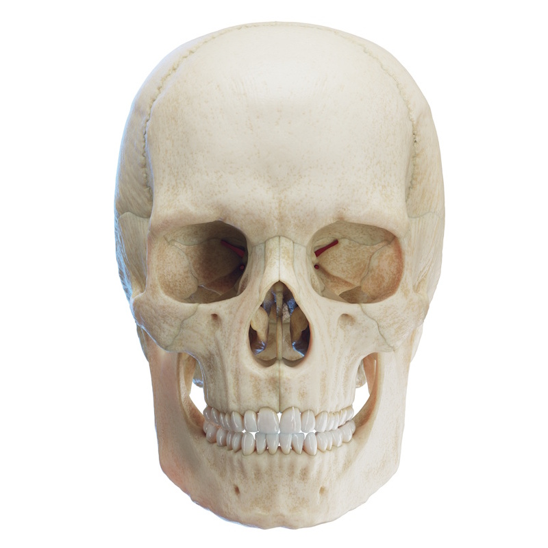
The ethmoid bone (os ethmoidale) is an unpaired bone situated centrally along the sagittal line of the skull between the two orbits, projecting inferior to the anterior cranial fossa (fossa cranii anterior). Belonging to the neurocranium, its name derives from the Greek word ethmos, meaning "sieve." The ethmoid bone contributes to the formation of the medial wall of the orbit, the nasal septum (septum nasi), the nasal and cranial cavities, and the floor of the anterior cranial fossa. It also serves a protective structural role, as the thin ethmoidal cells act as a buffer to absorb impact forces and protect the medial orbital wall from fracture.
The ethmoid bone consists of three main components:
- Cribriform plate (lamina cribrosa): A horizontal plate that forms the roof of the nasal cavity and part of the floor of the anterior cranial fossa. It transmits the fibers of the olfactory nerve (CN I).
- Perpendicular plate (lamina perpendicularis): A vertical plate extending inferiorly from the cribriform plate to form the upper two-thirds of the bony nasal septum.
- Ethmoidal labyrinth (labyrinthus ethmoidalis): Paired lateral parts situated on either side of the perpendicular plate, containing the ethmoidal air cells.
Articulations with Other Bones
Due to its central location, the ethmoid bone articulates with 13 other bones:
- Paired bones (10): Nasal bones (2), maxillas (2), lacrimal bones (2), palatine bones (2), inferior nasal conchae (2)
- Unpaired bones (3): The frontal bone, the sphenoid bone, the vomer
Cribriform Plate (Lamina cribrosa)
The cribriform plate is a horizontal structure seated within the ethmoidal notch (incisura ethmoidalis) of the frontal bone, forming a portion of the floor of the anterior cranial fossa. Its sieve-like architecture is perforated by numerous olfactory foramina (foramina cribrosa), through which the olfactory nerve fibers (fibrae nervi olfactorii / CN I) pass.
A triangular midline projection, the crista galli ("rooster's comb"), extends superiorly into the cranial cavity between the two olfactory bulbs to provide an attachment site for the falx cerebri (the vertical fold of dura mater separating the cerebral hemispheres). The anterior aspect of the crista galli expands into two lateral wings - the alae of the crista galli (alae cristae galli)- which articulate with the frontal bone.
Note: Some authors classify the crista galli as a superior extension of the perpendicular plate rather than an isolated component of the cribriform plate.
Perpendicular Plate (Lamina perpendicularis)
The perpendicular plate is a thin, flat bony sheet located in the median sagittal plane between the two ethmoidal labyrinths. Extending vertically downward from the cribriform plate, it forms the posterosuperior portion of the nasal septum, articulating anteriorly with the septal nasal cartilage (cartilago septi nasi) and inferiorly with the vomer.
Ethmoidal Labyrinth (Labyrinthus ethmoidalis)
The ethmoidal labyrinth consists of two lateral parts located on either side of the perpendicular plate, separating the orbit from the nasal cavity. It contains thin-walled, air-filled spaces known as the ethmoidal air cells (cellulae ethmoidales).
Anatomical features of the ethmoidal labyrinth:
- Ethmoidal air cells (cellulae ethmoidales): Divided into anterior, middle, and posterior groups (cellulae ethmoidales anteriores, mediae, et posteriores). They play an active role in vocal resonance (phonation) and respiration.
- Orbital plate (lamina papyracea): A paper-thin bony plate forming the lateral surface of the labyrinth and the medial wall of the orbit, covering the middle and posterior ethmoid cells.
- Superior nasal concha (concha nasalis superior): Forms the superior boundary of the superior nasal meatus.
- Middle nasal concha (concha nasalis media): Forms the superior boundary of the middle nasal meatus.
- Ethmoidal bulla (bulla ethmoidalis): A prominent, rounded bony elevation on the lateral wall of the middle meatus, formed by the expansion of the underlying anterior/middle ethmoidal air cells.
- Uncinate process (processus uncinatus): A delicate, sickle-shaped projection extending inferoposteriorly from the labyrinth to articulate with the ethmoidal process (processus ethmoidalis) of the inferior nasal concha.
- Ethmoidal infundibulum (infundibulum ethmoidale): A curved, slit-like air passage bounded medially by the uncinate process. It facilitates drainage for the maxillary sinus and anterior ethmoidal cells. The frontal sinus also drains into or near its anterior extension (frontal recess).
- Semilunar hiatus (hiatus semilunaris): A crescent-shaped groove located between the ethmoidal bulla superiorly and the uncinate process inferiorly. It serves as the primary communication and drainage channel into the middle meatus for the frontal, maxillary, and anterior ethmoidal sinuses.
Note: Occasionally, a small accessory concha - the supreme nasal concha (concha nasalis suprema) - is present superior and posterior to the superior nasal concha.