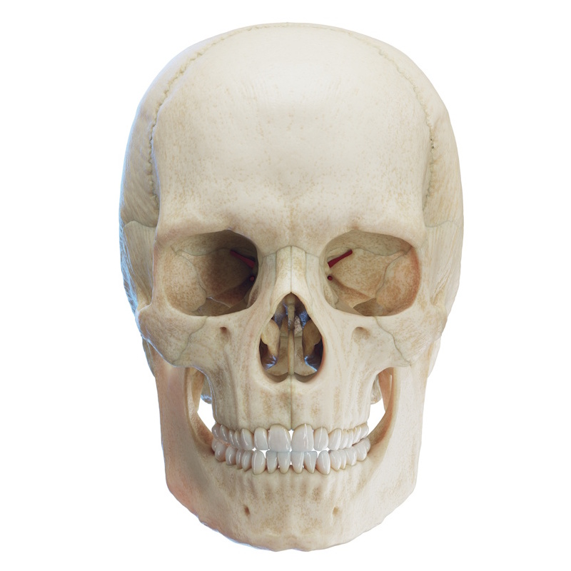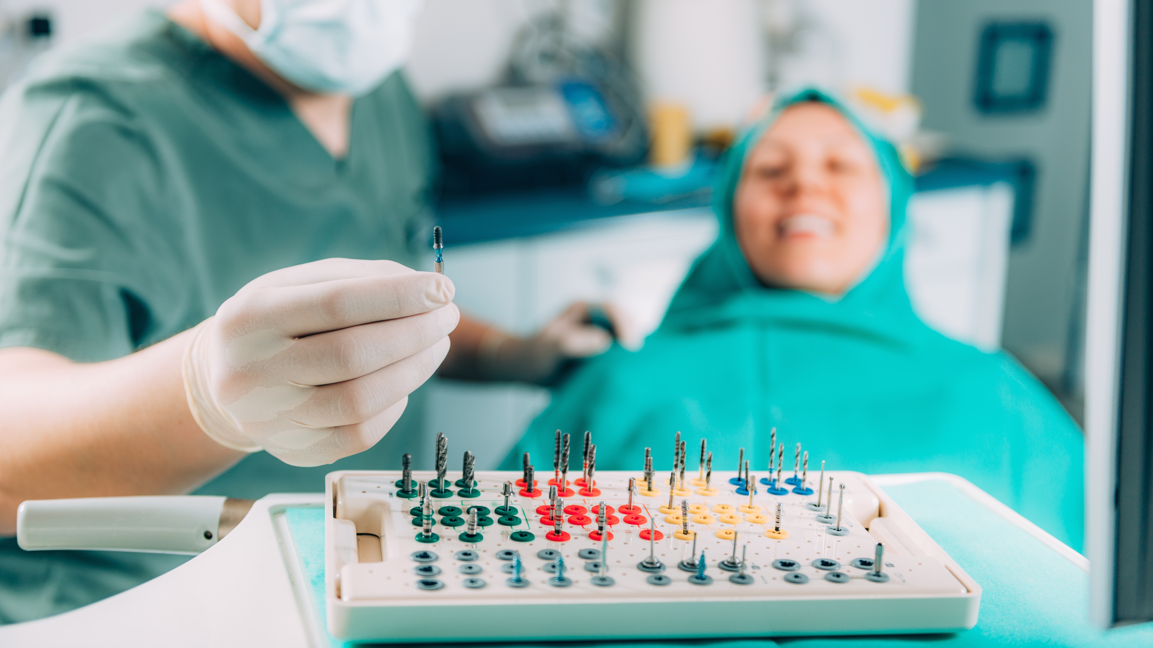

Information from CBCT imaging is essential for the proper planning of surgical guides, both for implant placement and other surgical procedures. In 90% of cases, we also need a virtual 3D model of the jaws, as well as preliminary digital modeling for the precise planning of the case. In the remaining 10% of cases, the surgical guide rests directly on the bone, relying entirely on the CBCT scan.
Below are our main recommendations for capturing a successful CBCT scan:
- Use High-End CBCT Scanners: Utilizing top-tier scanners yields optimal results.
- Scanning Volume / Field of View (FOV) Size: A high-end scanner does not always guarantee success. If the scanning FOV has a small diameter, the scanner's software is forced to perform more calculations, resulting in a less accurate synthesized image.
- Carefully Review Scan Results for Missing Data: Always check the scan for "cut-off teeth" or missing segments of the jaws being examined. The maxillary sinuses should be visualized at least halfway.
- Maintain Distance Between the Patient's Teeth: During the scan, patients should not close their teeth into full occlusion. Some authors recommend maintaining a 2 cm distance between the upper and lower dental arches. In most cases, having the patient bite on the machine's standard bite block (mouthpiece) is sufficient.
- Visualize the Vestibule: Capturing the outlines of the vestibule, lips, and cheeks during the CBCT scan is a crucial prerequisite for accurately adapting impressions to the scanner results. Two methods are most commonly used:
- Retraction: Pulling back the lips and cheeks using cotton rolls or an OptraGate is a good example.
- The "Pufferfish" Method: The patient bites on the mouthpiece, seals their lips tightly, and puffs out their cheeks. This pushes the cheeks and lips away from the teeth using the trapped air in the oral cavity.
- Patient's Tongue Position: When scanning the upper jaw, the tongue should be resting against the floor of the mouth. When scanning the lower jaw, the tip of the tongue should touch the soft palate. If scanning both jaws simultaneously, the specialist determines the tongue's position, but it is most commonly placed as it would be for a lower jaw scan.
- Always Scan Full Jaws: Regardless of the number of planned implants, always scan the entire jaw—never just a specific sector.
- Patient's Head Position: Centering the patient's head and eliminating any movement is of utmost importance for achieving optimal results.
- Remove All Removable Dentures: All removable prostheses must be taken out during the scan, unless you are creating a guide using a dual-scan protocol and radiographic markers have been placed on the dentures.
- Minimize Metal Artifacts: Dental crowns create "metal noise" (scatter) during scanning. Every manufacturer has built-in methods for reducing metal artifacts. If the artifacts cannot be reduced enough to properly adapt the patient's digital impression over the scan, a dual-scan protocol may be required.
- Exporting Scan Data: The scan data must always be exported in the universal .DICOM format.
- Pay Attention to Teeth with Compromised Periodontium: Teeth with periodontal disease may be highly mobile. Special care must be taken during the scan, as biting down on the mouthpiece could shift these teeth, making it impossible to accurately adapt the impression to them later.
Do you have any further questions? Do not hesitate to contact us!